
Fig. 2.
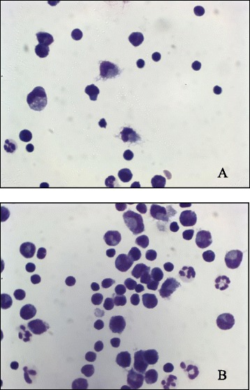
Cytological preparation of the cerebrospinal fluid of cases 2 (a) and 4 (b). a, b mononuclear pleocytosis. The cellular population was mainly represented by monocytes, macrophages, and lymphocytes. A small number of non degenerated neutrophils were also present (May-Grunwald Giemsa stain, ×100)