
Fig. 3.
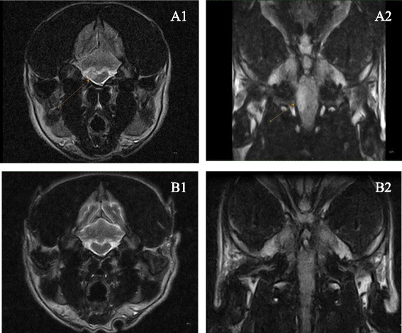
Dorsal and transversal MRI images of brainstem MOU lesion before and 6 months after treatment. The first MRI exam (A1 and A2) showed a single intra-axial hyperintense lesion (arrow) on the right side of brain stem, not too well-delineated, visible in T2-weighted sequences (A1) and FLAIR sequences (A2). There was no evidence of the lesion in the control MRI exam (B1 and B2) performed 6 months later