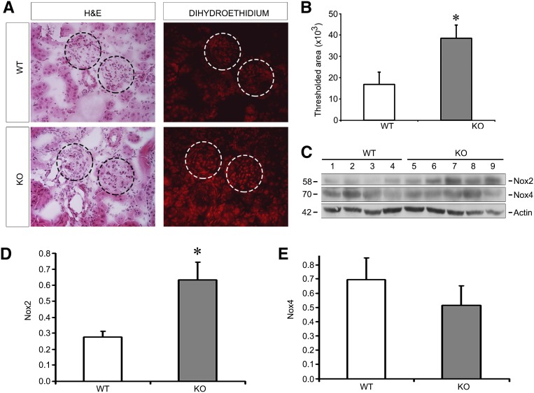
Figure 5.
Oxidative stress is increased in EC-SOD KO compared with WT mice treated with ADR. (A) DHE staining, which is specific for the superoxide free radical, is increased in KO versus WT mice. Circles outline glomeruli in these adjacent serial sections stained with H&E and with DHE. (B) Quantitation of positively stained area is shown. (C) Western blots for Nox2 and Nox4 reveal that Nox2 is higher in KO mice after ADR. (D and E) Quantitative data are shown in D and E normalizing to actin as a loading control. *P<0.05 compared with the WT group. DHE, dihydroethidium; H&E, hematoxylin and eosin.

