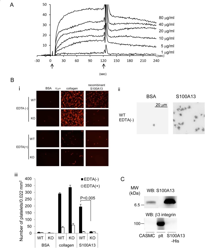
Fig 7. Recombinant S100A13 protein associated with CLEC-2.
A) Different concentrations of His-S100A13 were flowed over an immobilized hCLEC-2-rFc2 or a control rFc2-coated surface. The arrows indicate the beginning and the end of perfusion of S100A13. The results from one experiment are shown that is representative of the other three. RU indicates resonance units. B) (i) Platelets spreading on the surface of BSA, collagen, or S100A13 were investigated. (ii) Magnified images of adhered WT platelets on the surfaces coated with BSA or recombinant S100A13. (iii) Quantification of adherent platelets in the images in (i). Adherent platelets were counted. C) Western blotting with anti-S100A13 or anti-β3 integrin antibody. Plt represents platelets. The data are representative of three experiments.

