
Fig 10. S100A13 potentiated thrombus formation on collagen-coated surfaces under flow.
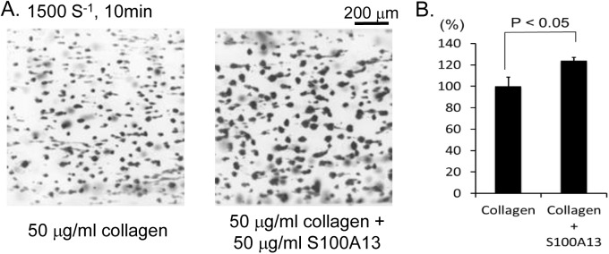
(A) Anti-coagulated human whole blood was perfused into cell microscopy chambers coated with collagen or collagen plus S100A13 for 10 min at a shear rate of 1500 s−1. Adherent platelets were visualized using a fluorescence video microscope. Movie data were converted into sequential photo images. (B) Thrombus volume was quantified. After 5 min of perfusion, adherent platelets were visualized by confocal laser microscopy, and the z-stack data were analyzed. The thrombus volume was expressed as the cIFI per image (404374 μm2). The graph illustrates the percentage of the surfaces coated with collagen only cIFI ± SE (n = 6, from two independent experiments).