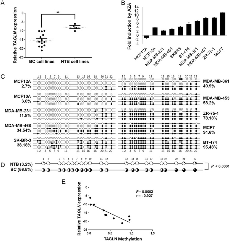
Fig. 2.

Expression and promoter methylation analyses of TAGLN gene in BC cell lines. a Box-plot analysis based on qRT-PCR results showing strong downregulation of TAGLN expression in BC cell lines compared to NTB cell lines. Log2 expression levels relative to GAPDH reference gene are shown. Horizontal dashed lines represent median values. Error bars: standard error of means (SEM). **P < 0.01; Mann–Whitney Test. b qRT-PCR analysis of AZA-treated BC and NTB cell lines indicating high levels of induction in TAGLN expression levels. Values represent the log2 ratios of expression levels in AZA-treated cell lines to those in their DMSO-treated cells. c QUMA software analyses based on bisulfite sequencing of BC and NTB cell lines showing hypermethylation in TAGLN promoter in BC cells. Percent values represent the average methylation of 5 clones for each cell line. Full circles: methylated; Empty circles: unmethylated. Sequence numbers of CpGs are shown above circles. d QUMA analysis showing significantly higher methylation in BC cells compared to NTB; Mann–Whitney test. Methylated DNA ratio: black area; unmethylated ratio: white area in the pie chart. e Scatter plot showing the spearman correlation of TAGLN methylation status with log2(expression) levels in BC and NTB cell lines
