Figure 4.
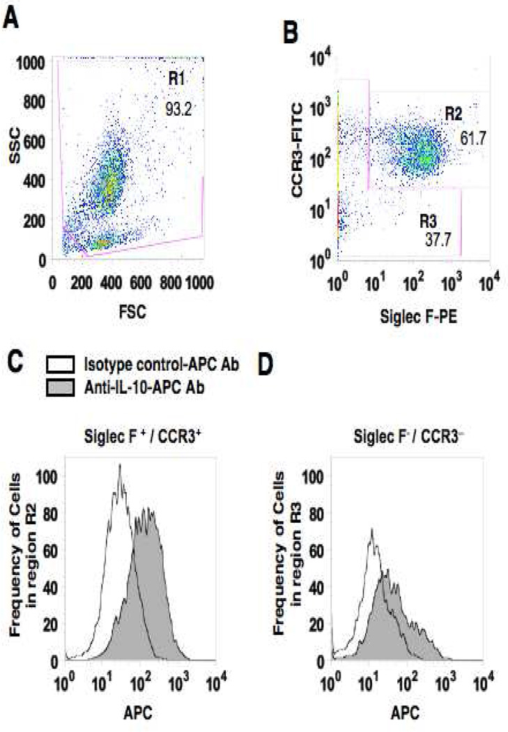
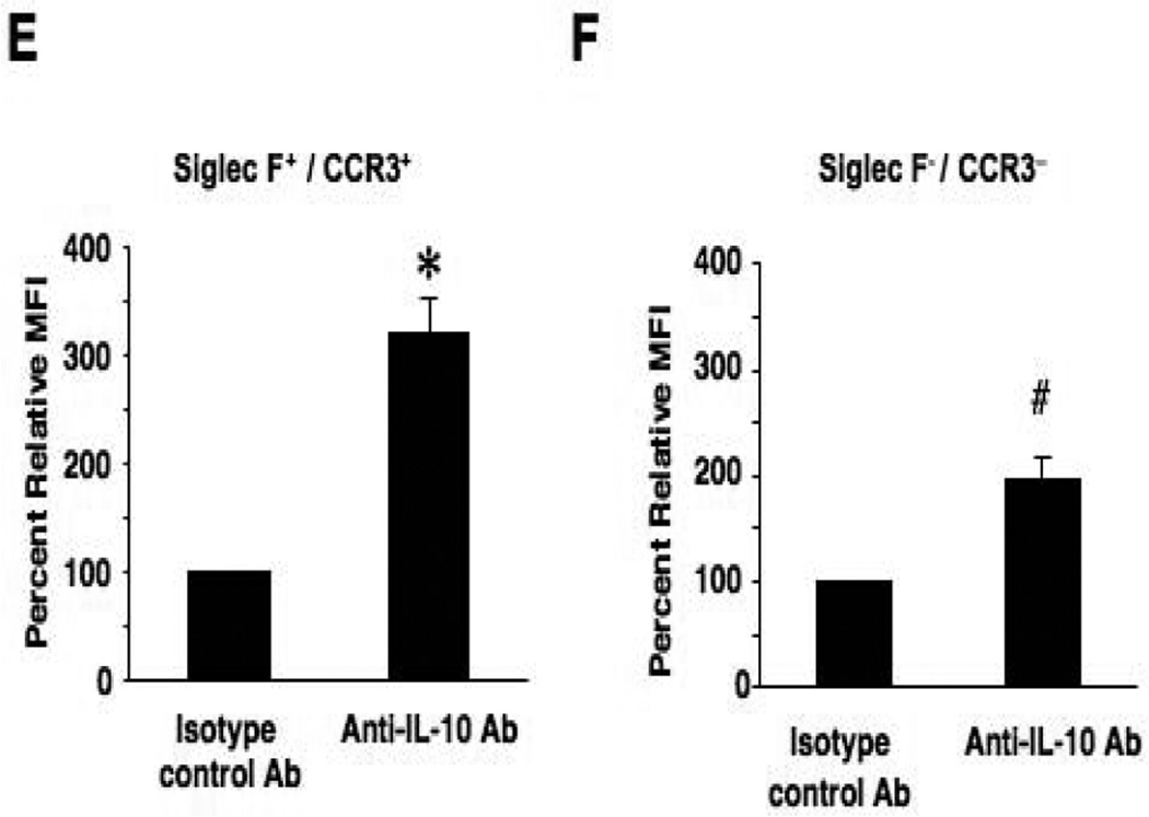
Intracellular IL-10 staining of airway eosinophils. BAL leukocytes from OVA sensitized and 7-challenged WT mice were activated with PMA/ionomysin and then stained with anti-mouse CCR3-FITC, anti-mouse Siglec-F-PE and anti-mouse IL-10-APC or APC-conjugated isotype control antibody. To identify the source of IL-10 in BAL fluid, live cells were first gated (region 1, R1) on forward scatter (FSC) and side scatter (SSC) (panel A). Cells in R1 were separated into Siglec-F- and CCR3-double-positive cells (R2) or negative cells (R3) (panel B). IL-10-positive cells in R2 or R3 were expressed as histograms (panel C and D) and the values for MFI were analyzed and expressed as the ratio to MFIs of isotype control staining in each sample (panels E and F). n=6. *p<0.001 and #p<0.01 vs. isotype control antibody group.


