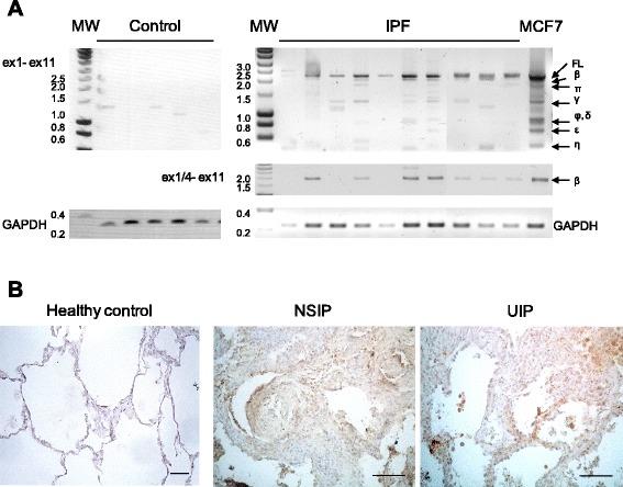
Fig. 6.

BARD1 expression is associated with human lung fibrosis. a RT-PCR with primers amplifying exon 1 to exon 11 (ex1- ex11) on tissues from control (non-symptomatic individuals) and a selection of IPF patients (total n = 17) is shown, performed with primers amplifying exon 1 to 11 or the region from exon 1/4 junction (BARD1β-specific) to exon 11 (ex1/4- ex11). Amplicons of BARD1 isoforms are indicated with Greek letters. GAPDH was amplified as control for RNA quality and quantity. In control lung tissues, only BARD1γ, BARD1δ, and BARD1η were detected, but no FL BARD1 or BARD1β. In tissues from fibrosis patients FL BARD1 and BARD1β were amplified in 70 percent of the samples. b Immunohistochemistry with anti-BARD1 antibody C20 shows representative cases of healthy lung tissue and tissues from patients with NSIP and UIP. Thickening interstitial areas and fibroblastic foci stained strongly for BARD1. Scale bars 100 μm
