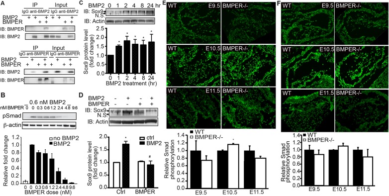
Fig 4. BMPER inhibits BMP2-induced signaling in the developing cushions.
(A) Recombinant BMPER and BMP2 proteins were combined as indicated and immunoprecipitated using an anti-BMPER antibody, an anti-BMP2 antibody, or the appropriate species-specific IgG antibody controls. As a loading control, all unbound proteins in the supernatant were run in the Input lanes (right lanes). The anti-BMP2 antibody recognized recombinant BMP2 and co-immunoprecipitated BMPER when both proteins were present. Similarly, the anti-BMPER antibody recognized recombinant BMPER and co-immunoprecipitated BMP2 when both proteins were present. (B) BMPER inhibits BMP2-induced Smad1,5,8 phosphorylation (pSmad) in cultured endothelial cells. MECs were treated with BMP2 and increasing doses of BMPER for 45 minutes. As expected, BMP2 treatment induced Smad phosphorylation (second lane). With increasing concentrations of BMPER, the pSmad levels were exponentially reduced (R2 = 0.98). Data are presented as the fold change compared with BMP2 treatment in the absence of BMPER treatment. Due to reduced signaling intensity when stripping and reprobing blots, β-actin was used as a loading control instead of total Smad. (C) BMP2 increases the Sox9 protein level in cultured endothelial cells. MECs were treated with 0.6 nM BMP2 for the indicated time periods. As expected, BMP2 treatment increased Sox9 protein levels. The arrow indicates the Sox9 protein band. N.S., not significant. *p<0.05, compared with cells without treatment. n = 3. (D) BMPER inhibits BMP2-induced Sox9 protein expression in cultured endothelial cells. MECs were treated with 0.6 nM BMP2 and 5 nM BMPER for 4 hours. BMPER co-treatment blocks BMP2-induced Sox9 protein expression. The arrow indicates the Sox9 protein band. N.S., not significant. *p<0.05, compared with control cells without treatment; #p<0.05, compared with cells with BMP2 treatment only. n = 3. (E) BMPER affects downstream Smad1/5/8 activity in the developing atrioventricular cushions. At E9.5, BMPER-/- atrioventricular cushions display reduced pSmad signals compared with their wild-type counterparts. However, by E10.5, the pSmad intensity increases in the BMPER-/- atrioventricular cushions compared with the wild-type counterparts. This increase is not maintained, with reduced pSmad intensity in the BMPER-/- cushions by E11.5. Fluorescence intensity is quantified on the right. (F) At E9.5, BMPER-/- outflow tract cushions display reduced pSmad compared with their wild-type counterparts. However, by E10.5, the pSmad intensity increases in the BMPER-/- outflow tract cushions compared with the wild-type counterparts. This increase is not maintained, with reduced pSmad intensity in the BMPER-/- cushions by E11.5. *p<0.05. Scale bar = 120 μm.

