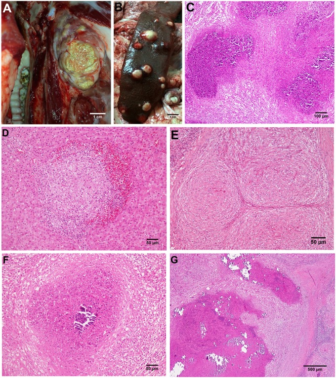
Fig 1. A-G.
A) TBL in the submandibular lymph node of an affected pig. Bar, 1cm. B) TBL in the spleen of an affected pig. Bar, 1cm. C) Microscopic image of a TBL lesions in the lymph node of an affected animal showing a profuse infiltrate of degenerated neutrophils. HE. Bar, 200μm. D) Clustered epithelioid macrophages surrounded by lymphocytes and erythrocytes in a stage I granuloma in the liver. HE. Bar, 50μm. E) Coalescent stage II granulomas in the lymph node of a pig showing epithelioid macrophages completely enclosed by a thin capsule, with peripheral infiltration of scattered lymphocytes. HE. Bar, 100μm. F) Stage III granuloma with a central necrotic core, partially mineralized, surrounded by a dense connective tissue capsule infiltrated by lymphocytes and scattered neutrophils. HE. Bar, 100μm. G) Thickly encapsulated, large, irregular, multicentric granulomas with prominent caseous necrosis and multifocal islands of mineralization (stage IV granulomas). HE. Bar, 500μm.

