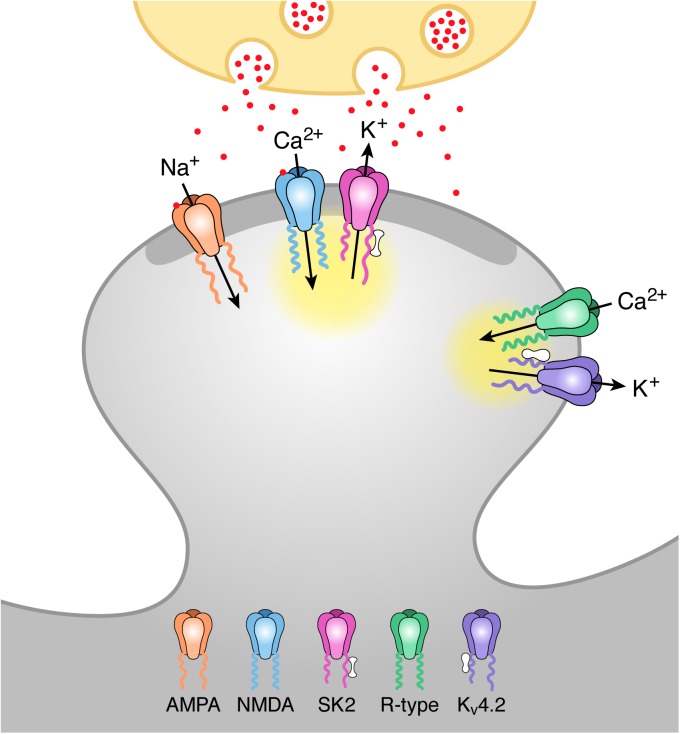
Fig 5. Model of activation of SK2 and KV4.2 containing channels by distinct Ca2+ microdomains during synaptic stimulation.
Schaffer collateral stimulation releases glutamate (red particles) from the presynaptic terminal (ivory). Glutamate binding to AMPA and NMDA receptors in the postsynaptic density (PSD; dark grey) of the spine head depolarizes the spine membrane potential and releases voltage-dependent Mg2+ block from NMDA receptors allowing for Ca2+ influx during the EPSP. This Ca2+ activates closely associated SK2 channels via binding to calmodulin (barbell structure) bound to C-terminus of SK2 subunits. Spine depolarization also activates R-type Ca2+ channels located extrasynaptically that are close to KV4.2-containing K+ channels. Ca2+ entering through R-type channels binds to KChIPs (peanut structure) associated with KV4.2 channels shifting the voltage-dependence of availability to more negative potentials and allowing for KV4.2 activation during an EPSP. The yellow clouds represent the microdomain for each Ca2+ source.

