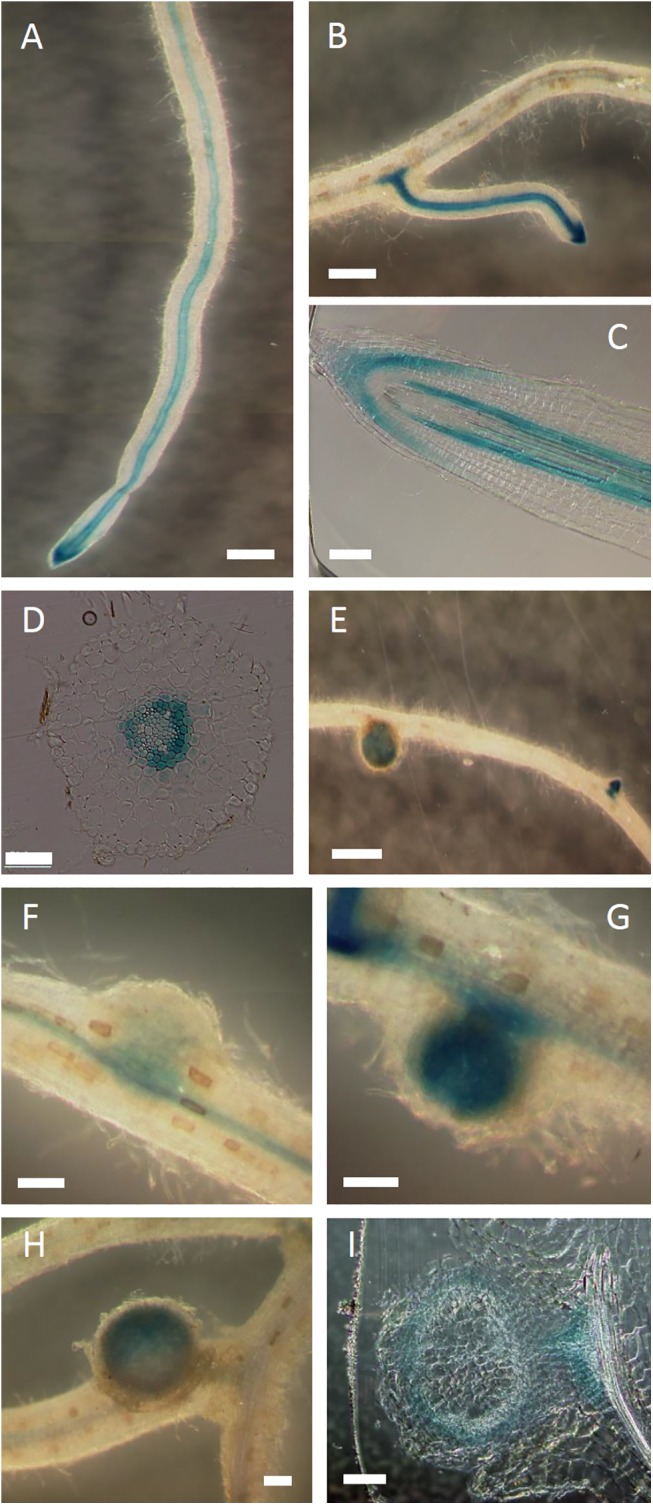
Fig 3. GUS staining of ProLjABCG1:GUS transformants infected with M. loti.

The transgenic hairy roots were stained with X-Gluc for 24 hours. (A, B) GUS activity in (A) roots and (B) lateral roots. (C) Longitudinal section of a root. (D) Cross-section of a root. (E) Root with a nodule and lateral root primordia. (F-H) Nodules of different stages from F (young) to H (mature). (I) Section of a nodule. Bars = 500 μm (A, B, E), 100 μm (C, F-I), and 50 μm (D).
