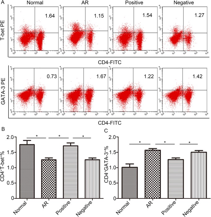
Fig 5. Lentiviral-mmu-miR-135a treatment influences Th cell polarization.
The expression of T-bet and GATA-3 protein in CD4+ T cells was measured in the spleens of normal (control), AR (AR-induced), positive (AR-induced, treated with lentiviral-mmu-miR-135a), and negative (AR-induced, treated with empty lentivirus) mice using flow cytometry. (A) Representative dot plots from each experimental group. The percentages of CD4+T-bet+ T cells (B) and CD4+GATA-3+ T cells (C) were also calculated. Data are presented as the mean ± SEM. *P<0.05.

