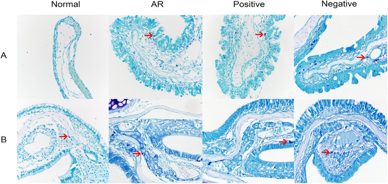
Fig 7. Mast cell staining of the nasal mucosa.

The nasal mucosa of mice in the normal (control), AR (AR-induced), positive (AR-induced, treated with lentiviral-mmu-miR-135a), and negative (AR-induced, treated with empty lentivirus) groups was analyzed 24 h after the previous OVA stimulation using MC staining. Representative images (original magnification: 400×) of the stained MCs (red arrows) present in sections of the nasal mucosa from each experimental group. A is the respiratory region of the nasal mucosa, B is the olfactory region of the nasal mucosa.
