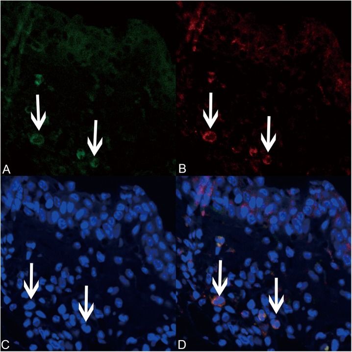
Fig 8. Lentiviral-mmu-miR-135a is expressed in mast cells.
Laser scanning confocal microscopy (original magnification × 400) was used to analyze the expression of lentiviral-mmu-miR-135a (green; A) and tryptase (red; B) in mast cells present in the nasal mucosa of the positive group. Cell nuclei were stained with DAPI (blue; C). The merged image (D) highlights the overlapped green and red signals around the mast cell nuclei.

