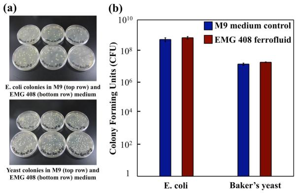
Figure 3.
Cell viability test of Escherichia coli and Saccharomyces cerevisiae. (a) Top and bottom photos show Escherichia coli and Yeast colonies formed in M9 medium and EMG 408 ferrofluids after 106 dilution from initial growth, respectively. (b) Colony Forming Unites (CFU) count of Escherichia coli and Saccharomyces cerevisiae using initial growth cell concentration.

