. 2015 Sep 21;16(3):297–306. doi: 10.4142/jvs.2015.16.3.297

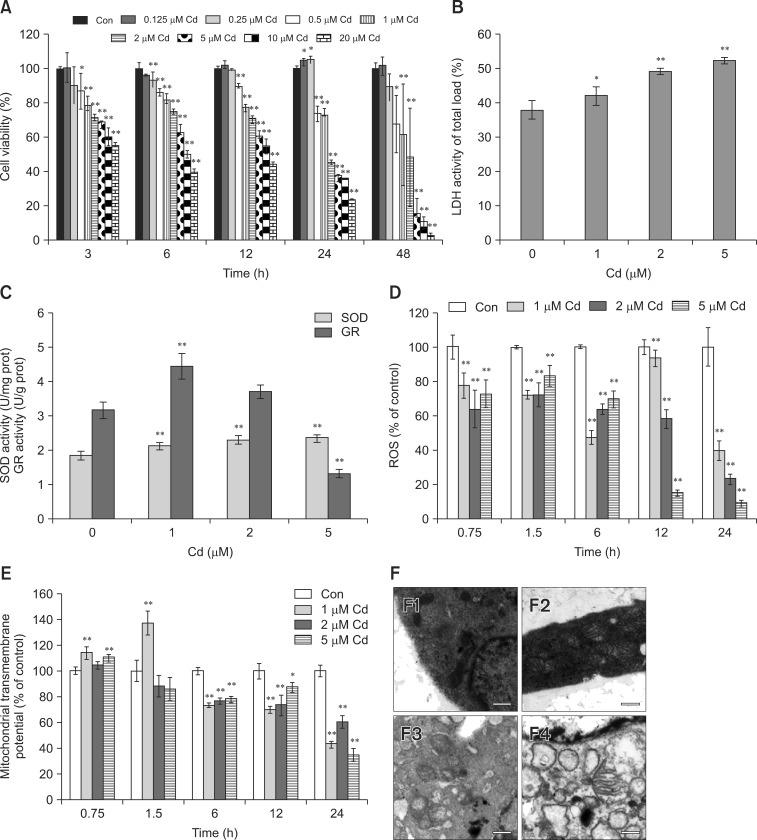
Fig. 1. Cadmium (Cd) altered cell viability, lactate dehydrogenase (LDH), antioxidant enzymatic activity, reactive oxygen species (ROS), mitochondrial membrane potential and ultrastructure in osteoblasts (OBs). (A) OBs were treated with 0-20 µM Cd, and cell viability was assessed at 3 to 48 h. (B and C) OBs were treated with 0-5 µM Cd for 24 h, and LDH, SOD, and glutathione reductase (GR) activities were determined. (D and E) Cells were exposed to 0-5 µM Cd for 0.75 to 24 h. Intracellular ROS production and mitochondrial transmembrane potential (MMP) were measured, and the results are presented as the mean fluorescence intensity (MFI) relative to the control group (100%). Data are presented as the mean and SDs of three independent experiments, each performed in triplicate. Significant differences are indicated by *p < 0.05 and **p < 0.01 relative to the control. (F) Ultrastructural morphology of OBs (5,200× magnification) after treatment with 0, 1, 2, and 5 µM Cd (F1-4) for 24 h.

Fig. 1