. 2015 Sep 21;16(3):297–306. doi: 10.4142/jvs.2015.16.3.297

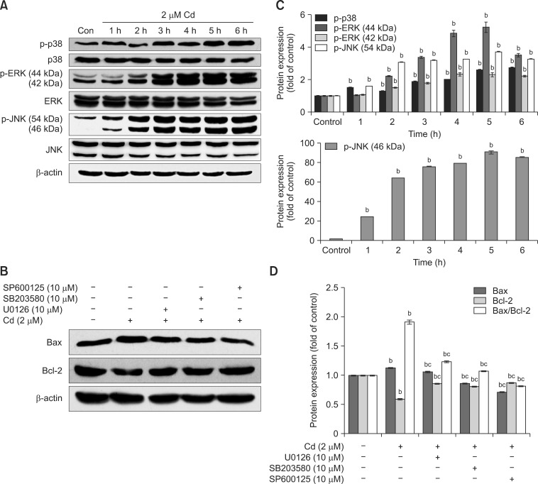
Fig. 4. The involvement of MAPK signaling in Cd-induced apoptosis. Cell lysates analyzed by western blot with the indicated antibodies. The β-actin protein level was used as a loading control. (A) Cells were cultured with 2 µM Cd for 1 to 6 h. (B) OBs were pre-incubated with 10 µM SB203580, SP600125, and U0126 for 30 min, followed by incubation with 2 µM Cd for 12 h. (C and D) The amount of phosphorylated protein was quantified by densitometry and corrected for sample loading based on the density of the β-actin band. The results are expressed as the fold changes relative to the control lane. Each blot is representative of at least three replicate experiments. ap < 0.05, bp < 0.01 relative to the control and cp < 0.01 relative to the Cd-treated group.

Fig. 4