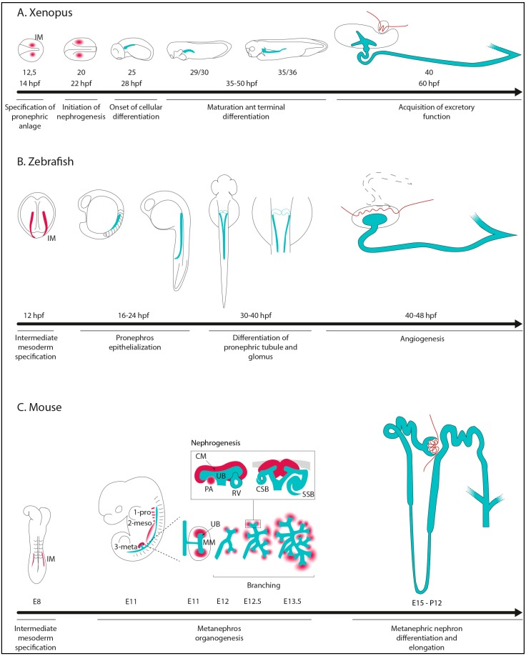
Figure 1.
Stages of kidney morphogenesis in Xenopus (A); zebrafish (B), and mouse (C). hpf: hours post-fertilization; IM: Intermediate Mesoderm; pro-: pronephros; meso-: mesonephros; meta-: metanephros; UB: Ureteric Bud; MM: Metanephric Mesenchyme; CM: Cap Mesenchyme; PA: Pretubular Agregate; RV: Renal Vesicle; CSB: Comma-Shaped-Body; SSB: S-Shaped-Body. IM, CM, and PA are shown in red, while renal epithelial tubular structures are in light blue.

