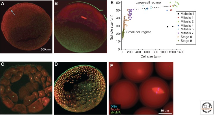
Figure 1.
Spindle-size scaling in Xenopus laevis. A–D show confocal images of eggs and early embryos fixed at different stages, stained for tubulin (red) and DNA (green), cleared and imaged by confocal microscopy. Embryos containing metaphase spindles were selected for analysis. (A) Unfertilized egg with meiosis-II spindle (blue arrow). (B) First mitosis. Note scaling mismatch between the spindle and egg. (C,D) Cleavage stages. (E) Spindle lengths and cell lengths derived from confocal images like A–D. Note spindle length is approximately constant in the large-cell regime and scales with cell size in the small-cell regime. (F) Spindle assembled in a droplet of unfertilized egg extract containing fluorescent probes suspended in oil and imaged live. aNuMA, anti-nuclear mitotic apparatus. (A–E from Wühr et al. 2008; adapted, with permission, from the author; F is an unpublished image provided by Jesse Gatlin, University of Wyoming, which is similar to images in Hazel et al. 2013.)

