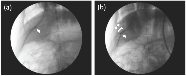
Figure 1.
Venography of the hepatic vein (HV) in the right medial lobe. (a) A small amount of contrast medium (CM) was injected from the proximal part of the HV, where a catheter tip was placed (arrow) to obtain a bird’s eye view of the HV in the right medial lobe. The HV was not occluded. (b) A fluoroscopic image of the injection of CM from the middle part of the HV (arrow, catheter tip) with a balloon occlusion (asterisks). CM was gently injected into the HV, and the peripheral branches were enhanced. The diameter of the HV, where the tip was placed in Figure 1b, was approximately 8 mm.

