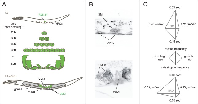
Figure 1.

MTs dynamics followed during differentiation in situ. (A) SM lineage. The left and right sex myoblasts (SML/R), divide and differentiated to generate 16 egg-laying muscles. UMCs = uterine muscle cells, VMCs = vulva muscle cells, VPCs = vulval precursor cells. (B) Maximal intensity projections of confocal z-series images of worms expressing GFP-tubulin under the control of the unc-62 promoter, at the L3 (top) and adult stages (bottom). (C) Diamond graphs representing dynamics data of MTs in the SM and UMCs. Data from Lacroix et al.12
