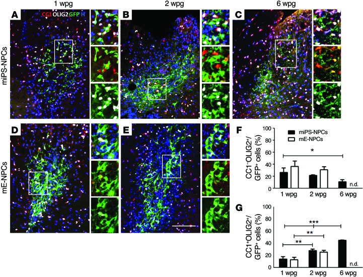
Figure 3. miPS-NPCs differentiate progressively into mature oligodendrocytes in response to demyelination in nude mice.
(A–E) Combined immunodetection of OLIG2 (white) and CC1 (red) with GFP (green) for miPS-NPCs (A–C) and mE-NPCs (D and E) at 1 (A and D), 2 (B and E), and 6 (C) wpg in myelin WT mice. (F and G) Quantification of CC1–/OLIG2+ (F) and CC1+/OLIG2+ GFP+ cells (G). The percentage of immature oligodendrocytes decreased significantly after 2 wpg for both grafted cell types, while the percentage of mature oligodendrocytes increased for both miPS-NPCs and mE-NPCs. One-way analysis of variance plus Tukey’s multiple comparison tests and Student’s t test were used for the statistical analysis of these experiments (n = 3–4 mice per group). *P < 0.05, **P < 0.01, and ***P < 0.001. H: Hoechst dye; n.d.: not done. Scale bar: 100 μm.

