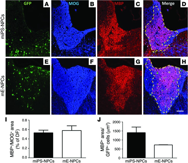
Figure 6. Competitive remyelination potential of iPS-derived cells in demyelinated Shi/Shi Rag2–/– mice.
(A–H) Colabeling of MOG (blue) to detect whole myelin with MBP (red) to detect myelin derived from exogenous cells on coronal sections of demyelinated Shi/Shi Rag2–/– spinal cord grafted with miPS-NPCs (A–D) and mE-NPCs (E–H) reveals extensive remyelination by exogenous cells compared with endogenous cells. (I) No statistical difference between miPS-NPC– and mE-NPC–derived myelin was detected 6 wpg. (J) When the amount of myelin was expressed per number of GFP+ cells in each transplanted group, the myelination domain of miPS-NPCs was greater than that of mE-NPCs, but not statistically different. Dotted lines represent the MOG+/MBP+-labeled area. Mann Whitney test was used for statistical analysis (n = 3–4 mice per group). Scale bar: 50 μm.

