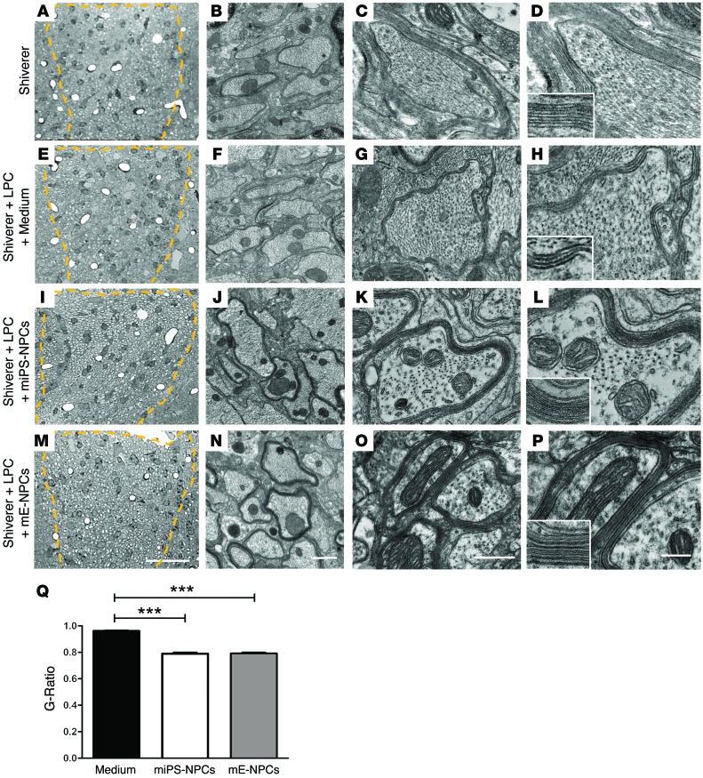
Figure 8. miPS-derived oligodendrocytes produce compact myelin in the demyelinated Shi/Shi Rag2–/– spinal cord.
(A–P) Ultrastructure of myelin on coronal sections of the dorsal funiculus (A–D) of intact Shi/Shi Rag2–/– mouse spinal cord; (E–H) 6 weeks after LPC-induced demyelination followed by medium injection; (I–P) 6 weeks after LPC injection and engraftment of miPS-NPCs (J–L) and mE-NPCs (M–P). While Shi/Shi Rag2–/– axons were surrounded by wraps of loose uncompacted myelin in normal or remyelinated (with fewer warps) conditions, they were surrounded by thick and compact normal myelin derived from the grafted NPCs (identified at G–L for miPS-NPCs and N–P for mE-NPCs). No difference in compaction and structure is observed between myelin derived from miPS-NPCs and mE-NPCs. (Q) Quantification of g ratio revealed a significant difference between myelin thickness of axons remyelinated by endogenous cells versus that of axons remyelinated by miPS-NPCs or mE-NPCs. Dotted lines in A, E, I, and M represent the lesion site in the dorsal funiculus. One-way analysis of variance plus Tukey’s multiple comparison tests were used for graph Q (n = 3–4 mice per group). Insets in D, H, L, and P are enlargements of myelin, illustrating its fine structure. ***P < 0.001. Scale bars: 30 μm in A, E, I, and M; 1 μm in B, F, G, and N; 500 nm in C, G, K, and O; and 200 nm in D, H, L, and P.

