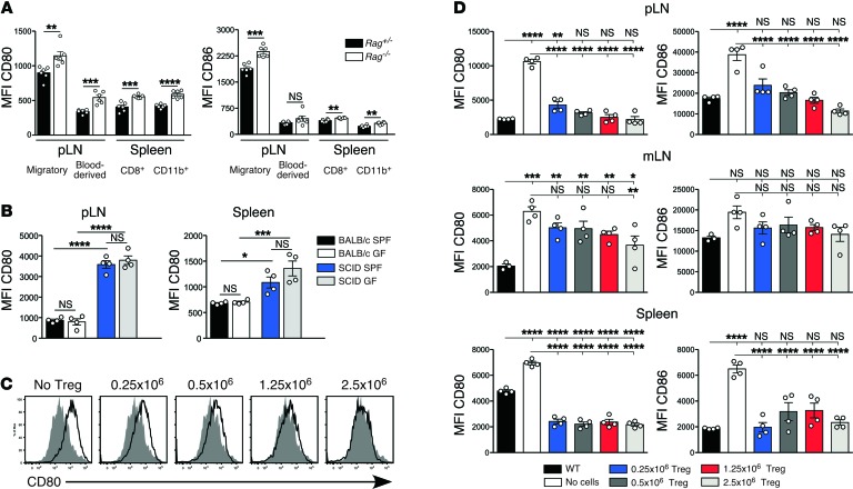
Figure 2. Expression of CD80 and CD86 by DCs from Treg-deficient mice.
(A) MFI of CD80 (left panel) and CD86 expression (right panel) by DCs from immunodeficient Rag–/– mice and their immunosufficient Rag+/– littermates (n = 6 per group). Migratory DCs and blood-borne DCs in pLN/mLN were gated as MHCIIhiCD11cint and MHCIIintCD11chi, respectively. Blood-borne DCs in the spleen were further subdivided based on expression of CD8 and CD11b. Results are from a single experiment and are representative of more than 20 independent experiments. (B) Comparison of DC phenotype in mice housed under SPF versus GF conditions. MFI of CD80 expression by CD11c+B220– pLN and spleen DCs from immunosufficient BALB/c and immunodeficient C.B-17/lcr-scid mice housed under either SPF or GF conditions. Data are pooled from 2 independent experiments with 2 mice per group in each experiment. (C) Expression of CD80 by migratory DCs in the pLN of Rag–/– mice that received either no cells or were reconstituted with 0.25 × 10⁶, 0.5 × 10⁶, 1.25 × 10⁶, or 2.5 × 10⁶ CD4+CD25+ Tregs (open histograms) compared with expression in untreated WT mice (shaded histograms). (D) Expression of CD80 (left panels) and CD86 (right panels) by migratory DCs in the pLN, mLN, and splenic DCs at day 7 after Treg reconstitution compared with expression in untreated WT and Rag–/– mice. Data in C and D are from a single experiment with n = 4 per group. The comparison of WT, Rag–/– mice, and Rag–/– mice reconstituted with 2.5 × 10⁶ CD4+CD25+ Tregs is representative of more than 10 independent experiments. Data in A were analyzed using a 2-tailed t test, while data in B and D were analyzed using one-way ANOVA with a Newman-Keuls post-test. Bars represent mean ± SEM with individual values indicated by the open circles. *P < 0.05, **P < 0.01, ***P < 0.001, and ****P < 0.0001.

