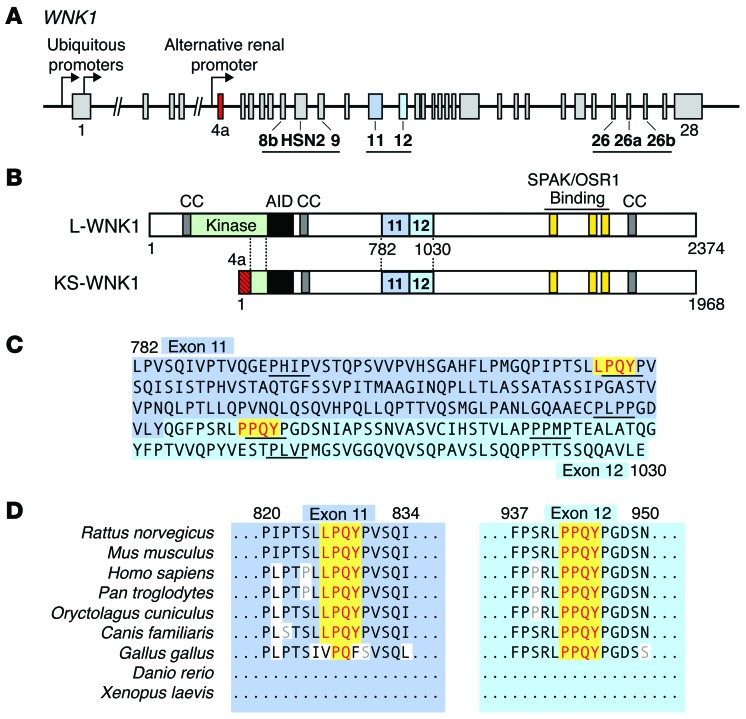
Figure 1. Identification of 2 PY motifs in exons 11 and 12 of WNK1.
(A) Schematic representation of the WNK1 gene. Two ubiquitous promoters drive the transcription of WNK1 isoforms containing an intact kinase domain (L-WNK1). A renal promoter drives the expression of a kidney-specific transcript (KS-WNK1). Regions of WNK1 that undergo tissue-specific alternative splicing are underlined. Exon 4a, which encodes a short N-terminal sequence unique to KS-WNK1, is shown in red. Exons 11 and 12 are highlighted in blue. (B) Domain structure of L-WNK and KS-WNK1. The location of 3 coiled coil domains (CC) that facilitate WNK complex formation (65), an autoinhibitory domain (AID) that suppresses WNK kinase activity (66), and C-terminal SPAK/OSR1 binding motifs are shown. Downstream of exon 4a, L-WNK1 and KS-WNK1 are identical. (C) Amino acid sequence of rat exons 11 and 12. Two canonical PY motifs are highlighted in yellow. The exons are proline rich and contain numerous PXXP motifs. (D) The PY motifs in exons 11 and 12 are highly conserved in mammals. The exons are not present in nonmammalian organisms, such as Xenopus, zebrafish, Drosophila, and C. elegans.

