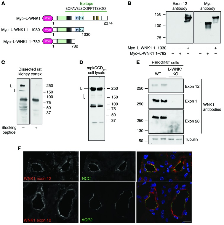
Figure 2. WNK1 isoforms containing PY motifs are expressed in the ASDN.
(A) Schematic representation of N-terminally Myc-tagged cDNAs used for exon 12 antibody validation. Rabbit antisera to WNK1 were generated to the indicated exon 12 epitope. (B) The exon 12 antibody specifically recognizes a Myc-tagged L-WNK1 fragment containing exon 12 that was transiently expressed in HEK-293T cells. (C) Immunoblots of dissected rat kidney cortex homogenates (50 μg) probed with the exon 12 antibody revealed a discrete band at approximately 250 kDa, corresponding to the MW of L-WNK1 (L), and shorter species migrating between 150 and 250 kDa, consistent with KS-WNK1 and/or C-terminal proteolytic fragments of L-WNK1 (bracket). Both high-MW bands were not seen when the antibody was preincubated with excess immunizing peptide. Several lower-MW species were also quenched with peptide competition. A nonspecific band was noted between 75 and 100 kDa. (D) Bands of similar MWs were noted in mpkCCDc14 CCD cells. (E) Along with 2 other WNK1-specific antibodies, the antisera recognized a band at approximately 250 kDa in HEK-293T cells that was absent in genetically validated L-WNK1 KO cells (19). (F) Immunostaining of mouse kidney with exon 12 antisera revealed a tubule-specific signal that colocalized with NCC in DCT and aquaporin-2 (AQP2) in CCD. Scale bars: 10 μm. See also Supplemental Figures 1 and 2.

