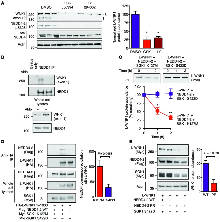
Figure 6. SGK1 attenuates NEDD4-2–mediated WNK1 degradation.
(A) Left: mpkCCDc14 cells were pretreated with GSK650394 (10 μM), LY294002 (50 μM), or DMSO. Two hours later, cells were stimulated with 100 nM aldosterone for 5 hours and whole cell lysates (20 μg) were subjected to SDS-PAGE and immunoblotting with the indicated antibodies. High-MW signals at approximately 250 kDa, corresponding to L-WNK1 (L) and lower-MW species corresponding to KS-WNK1 and/or proteolytic fragments of L-WNK1 (bracket) are noted. Right: quantification of L-WNK1 abundance. n = 3; P < 0.01 for either drug vs. DMSO control by 1-way ANOVA, Dunnett’s test. (B) Co-IP assay in mpkCCDc14 cells. Anti–Pan-NEDD4 immunoprecipitates (targeting both NEDD4-1 and NEDD4-2) from cells pretreated with 100 nM aldosterone or vehicle for 14 hours. Representative of 3 experiments. (C) HEK-293T cells expressing full-length Myc-tagged L-WNK1 (2 μg/well) and NEDD4-2 (1 μg/well) in the presence of either kinase active SGK1-S422D or dead SGK1-K127M (1 μg/well) were chased with 100 μg CHX for 2 hours. n = 4; P < 0.05 at 1 hour and P < 0.01 at 2 hours by Student’s t test. (D) Co-IP assay in HEK-293 cells. Anti-HA immunoprecipitates of L-WNK1 1-1030 with NEDD4-2 in the presence of either active (S422D) or kinase dead (K127M) SGK1. n = 4; P = 0.0458 by Student’s t test. (E) Steady-state abundance of L-WNK1 transiently expressed in HEK-293T cells with SGK1 S422D and either WT NEDD4-2 or phosphorylation-resistant (PR; S338A/T363A) NEDD4-2. n = 4; P = 0.007 by Student’s t test.

