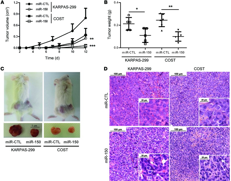
Figure 6. miR-150 overexpression inhibits KARPAS-299 and COST xenograft growth in NOD/SCID mice.
(A) NPM-ALK(+) KARPAS-299 and COST cells transfected either with miR-CTL or miR-150 were injected s.c. in the left or right flank of 5 NOD/SCID mice, respectively (n = 5). Tumor volume was evaluated over time by caliper measurements and reported as mean ± SEM (bars). **P < 0.001, ***P < 0.0001, using unpaired 2-tailed Student’s t test. (B) Tumor weight measurement, reported as mean ± SEM (bars). *P < 0.05, **P < 0.001, using unpaired 2-tailed Student’s t test. (C) Representative tumors resected from mice xenografted with miR-CTL– or miR-150–transfected KARPAS-299 and COST cells. Scale: 1 cm. (D) Micrographs of hematoxylin and eosin staining of excised miR-CTL or miR-150 tumors (scale bars: 100 μm, inset 20 μm; original magnification ×20, inset ×80). Arrows indicate cells with phenotypic hallmarks of cellular degeneration, i.e., uncommon chromatin condensation, nuclear piknosis, cellular volume decrease, or nuclear envelope disruption.

