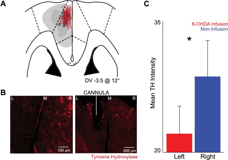
Fig. 2.
Infusions of 6-OHDA in rodent MFC causes dopamine depletion. A: histology from brain slices from 8 animals revealed approximate electrode placement in the MFC (red dots). Cannula from all 11 animals located within MFC and within recording distance of the electrode tips (blue dots) are shown. Light gray patch, approximate maximum region of dopamine depletion; dark gray path, approximate minimum region of dopamine depletion. DV, dorsal-ventral. B: low and medium power coronal sections stained with tyrosine hydroxylase (TH; red); MFC 6-OHDA was injected on the left side. M, midline; R, right; L, left. C: there was less TH+ staining in sections ipsilateral to MFC 6-OHDA infusion (left) compared with control, contralateral sides without infusion (right). Values are means ± SE. *Significance at P < 0.05 via paired t-test.

