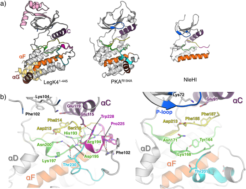
Figure 3. Structure of apo-LegK41–445.
(a) Cartoon representation of the crystal structures of apo-LegK41–445, mouse PKA mutant R194A (pdb code 4DFY) and NleH1 (pdb code 4LRJ). The helices αC, αF and αG are coloured in purple, orange and brown, respectively. The P- loop is coloured in blue and the activation segment coloured as follow: catalytic loop (green), Magnesium-binding loop (yellow), activation loop (magenta) and P+1 loop (cyan). (b) Close-up view of the catalytic site of apo-LegK41–445 (left) and PKA mutant (right) coloured as in (a). A side chain of important residues are shown as ball and sticks and coloured as the cartoon (carbon), red (oxygen) and blue (nitrogen). Main hydrogen bonds are indicated as dashed lines.

