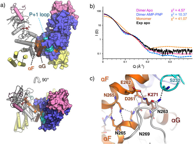
Figure 5. LegK41–445 dimer organisation.
(a) Side (upper panel) and top (lower panel) views of the dimer with chain A coloured in grey and shown as a surface and chain B coloured in blue and shown as ribbon. The cap and FHB domains of the two chains are coloured in pink and yellow, respectively. (b) Comparison of the experimental SAXS curve of apo-LegK41–445 (black) with theoretical SAXS curves of apo-LegK41–445 monomer (orange), dimer (magenta) and AMP-PNP•LegK41–445 dimer (blue). χ2 obtained with FOXS server51 are indicated. (c) Detailed view of the dimer interface coloured as in (a) with participating residues shown as ball and stick.

