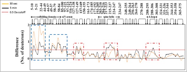
Figure 1.

Difference in deuterium exchange between protease-free procapsids and protease containing procapsids at peptide resolution highlight the direct and indirect effects of protease binding. Specific pepsin-digest fragment peptides of procapsid protein generated are listed from the N- to C-terminus (x-axis). The 2 plots represent the difference in deuterium exchange (y-axis) at 2 time points of deuterium on-labeling (t = 30 sec (orange) and t = 5 min (black)) with each point representing a single pepsin-digest fragment peptide. Positive differences represent regions in the procapsid showing reduced deuterium exchange in presence of protease. Peptides showing differences at early time points represent direct binding sites (blue dashed boxes) and those regions showing larger differences in deuterium exchange (t = 5 min) represent long range stabilization/allosteric sites (red dashed boxes). Deuterium exchange of 0.5 Da is considered significant and is represented by a red dashed line in the plot.
