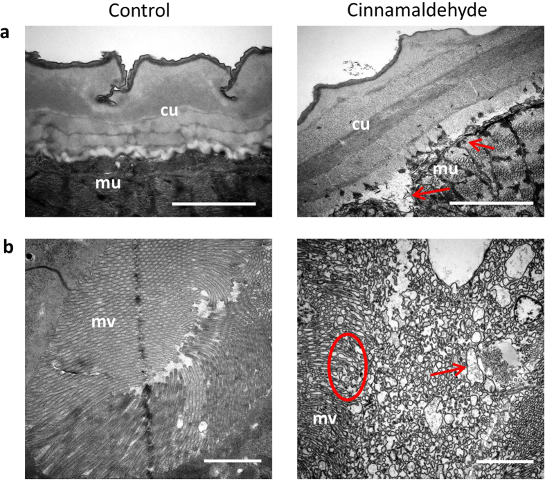
Figure 6. Ultrastructural changes in Ascaris suum exposed to trans-cinnamaldehyde.
Transmission electron micrographs of A. suum fourth-stage larvae exposed to either culture media (Control) or 236 μM trans-cinnamaldehyde (CA) for 12 hours. For all panels scale bar indicates 2 μm. (a) Cuticle (cu) and underlying Muscular (mu) tissue—note the lesions in the muscle tissue underlying the cuticle and hypodermis in parasites exposed to CA (red arrows). (b) Digestive tissues showing the microvilli (mv) overlying the intestinal lumen—note the destruction of the villi (red circle) and the presence of large vacuoles (red arrow) in parasites exposed to CA.

