
Fig. 1.
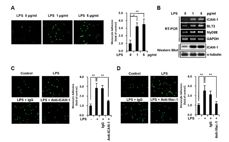
ICAM-1 expression induced by LPS in MDA-MB-231 breast cancer cells promotes their adhesion to THP-1 monocytes. (A) MDA-MB-231 cells were treated with LPS (0, 1, and 5 μg/ml) for 24 h and then co-cultured with calcein-AM-labeled THP-1 cells for 1 h. THP-1 cells that had adhered to MDA-MB-231 cells were visualized, and the number of adherent monocytes was determined using fluorescence microscopy. (B) MDA-MB-231 cells were treated with LPS (0, 1, and 5 μg/ml) for 24 h, after which the mRNA levels of ICAM-1, BLT2, MyD88 and GAPDH were measured by semi-quantitative RT-PCR. The protein levels of ICAM-1 and α-tubulin were measured by Western blot assay. (C) LPS-pretreated MDA-MB-231 cells (1 μg/ml, 24 h) were treated with anti-human ICAM-1 or anti-mouse IgG isotype control antibodies for 2 h, and the cells were then co-cultured with calcein-AM-labeled THP-1 cells for 1 h. The adherence of THP-1 cells to MDA-MB-231 cells was visualized, and the number of adherent monocytes was determined using fluorescence microscopy. (D) THP-1 cells were treated with anti-human Mac-1 or anti-mouse IgG isotype control antibodies for 2 h, and the THP-1 cells with calcein-AM-label were then co-cultured with LPS-pretreated MDA-MB-231 cells for 1 h. The adherence of THP-1 cells to MDA-MB-231 cells was visualized, and the number of adherent monocytes was determined using fluorescence microscopy. The data are representative of three independent experiments with similar results. All quantitative data are represented as the mean ± SD from three independent experiments. *p < 0.05, **p < 0.01.