Figure 3.
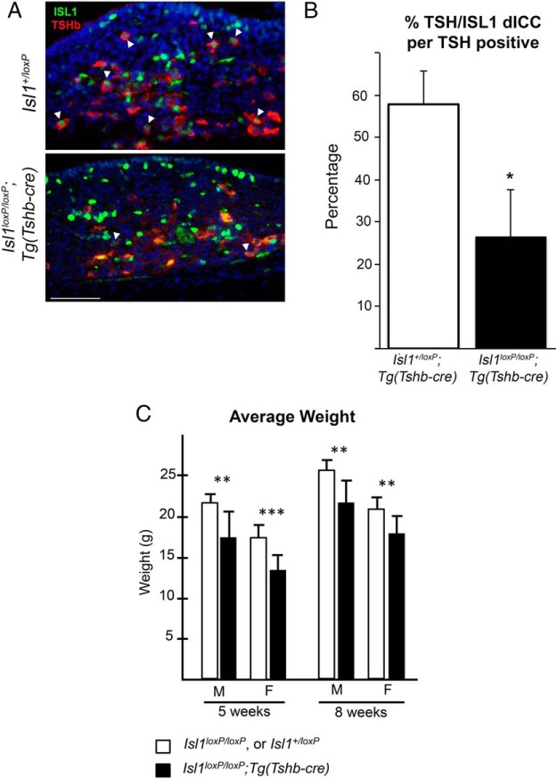
Isl1loxP/loxP;Tg(Tshb-cre) mice present a significant growth insufficiency. A, Coronal pituitary sections from P3 Isl1loxP/loxP;Tg(Tshb-cre) and control mice were immunostained with ISL1 (green nuclear stain) and TSH (red cytoplasmic stain). Cells with both ISL1 and TSH are present in the control sections (white arrowheads). There are very few cells with ISL1 and TSH in the Isl1loxP/loxP;Tg(Tshb-cre) mice (white arrowheads). Photos were taken at ×630. Scale bar, 50 μm. B, The percentage of TSH cells that also express ISL1 (dICC, double immunopositive) was calculated for the Isl1loxP/loxP;Tg(Tshb-cre) mutant and control mice. The mutant mice have significantly fewer colocalized ISL1+, TSH+ cells in their pituitaries compared with controls (*, P < .05). C, Average weights of male and female Isl1loxP/loxP;Tg(Tshb-cre) and control mice at 5 and 8 weeks. There is a significant difference between both male and females at both ages. At 5 weeks, Isl1loxP/loxP;Tg(Tshb-cre) males are 17.2 ± 3.2 g (n = 9), control males are 21.8 ± 1.0 g (n = 8), Isl1loxP/loxP;Tg(Tsh-bcre) females are 13.5 ± 1.6 g (n = 10), and control females are 17.2 ± 1.5 g (n = 14). At 8 weeks, Isl1loxP/loxP;Tg(Tshb-cre) males are 21.9 ± 2.6 g (n = 6), control males are 25.6 ± 1.4 g (n = 10), Isl1loxP/loxP;Tg(Tshb-cre), and control females are 20.9 ± 1.5 g (n = 10). Statistical significance: ***, P < .001; **, P < .01.

