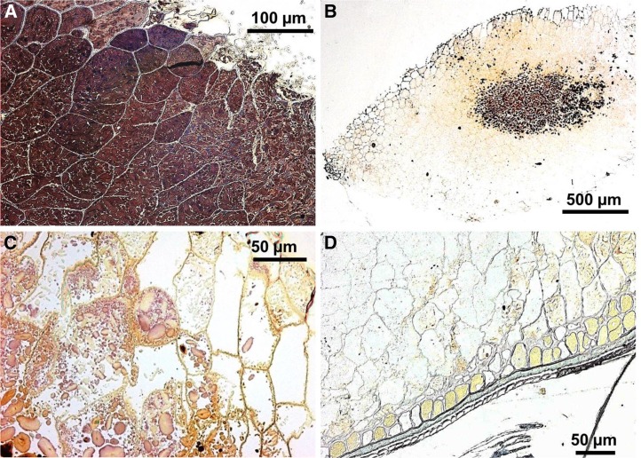
FIGURE 5 .
Sections of wheat endosperm particles from coarse porridge at various stages of digestion. All panels are light microscopic micrographs in which the starch was stained with 2.5% (wt:vol) Lugol’s iodine. Starch-filled wheat endosperm tissue from the cooked coarse porridge before digestion (A). Low-magnification view of a typical 2-mm wheat particle recovered in ileal effluent after 4 h (B). The staining pattern suggests a progressive digestion of starch from the particle periphery toward the core. A higher-magnification view than in panel B shows the digested edge of a particle collected from effluent after 4 h (C). Starch in the outermost cell layers (toward the right) has been digested, leaving empty cells. Typical particle remnant recovered in ileal effluent during the night (22-h gut residence time) (D). In the overnight samples, remnants of endosperm tissue were observed only when attached to adjacent outer tissue layers (aleurone, pericarp, and testa), and most of the starch had been digested.

