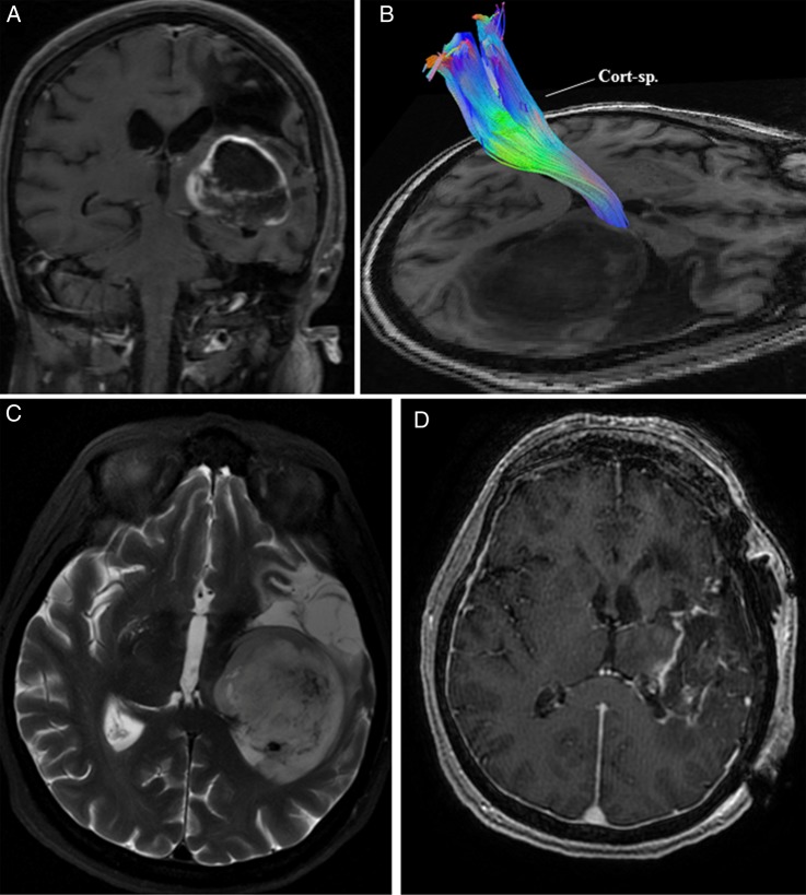
Fig. 2.
(A) MRI of the brain in Case 2 demonstrated a left-sided contrast-enhancing basal ganglia mass with evidence of a previous middle cerebral artery territory infarction. (B) Preoperative high-definition fiber tractography revealed a displaced corticospinal tract (Cort-sp.) in the perilesional area. (C) T2-weighted MRI of the brain further delineated the tumor. (D) Postoperative contrast-enhanced MRI showed a residual cuff of tumor along the margin adjacent to the corticospinal tract and thalamus.

