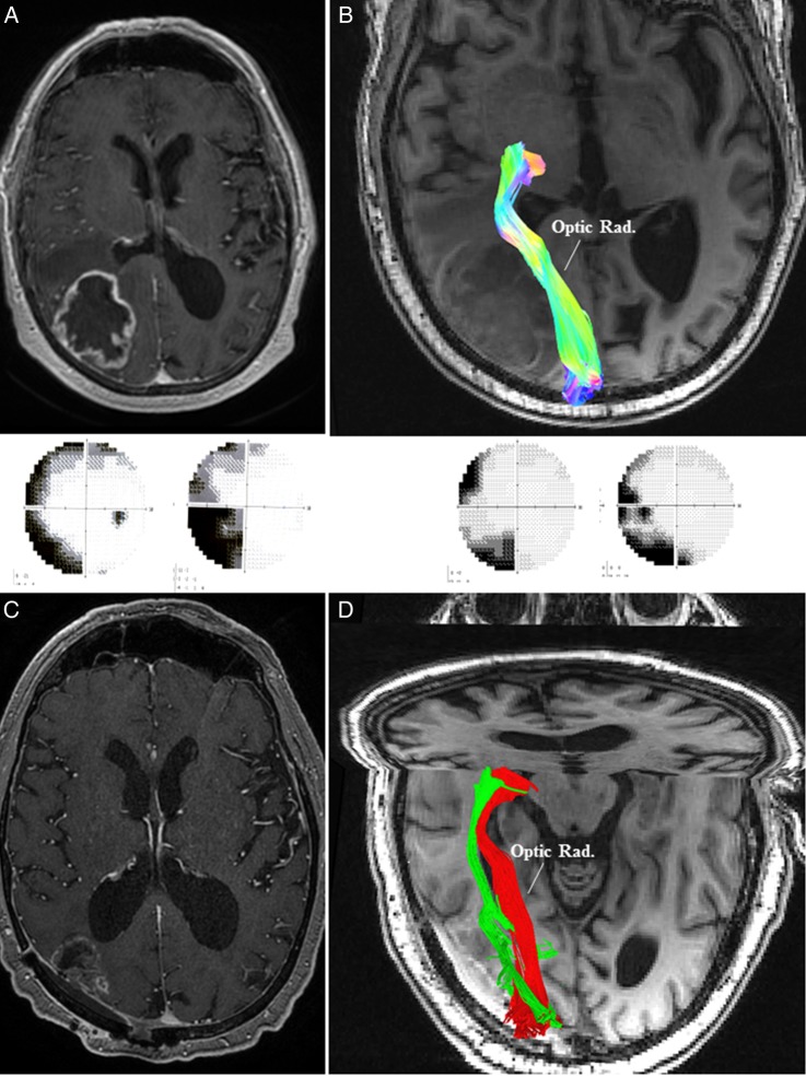
Fig. 3.
(A) MRI of the brain in Case 3 demonstrated a right-sided contrast enhancing parieto-occipital lesion leading to left-sided homonymous hemianopia (panel below). (B) Preoperative high-definition fiber tractography (HDFT) showed medially and inferiorly displaced optic radiation (Optic Rad.) in the perilesional edematous area. (C) Postoperative MRI with contrast confirmed an adequate resection with a small residual on the medial margin adjacent to the optic radiation. (D) Postoperative HDFT confirmed the improvement in the configuration of the displaced optic radiation (red: preoperative; green: postoperative). This was accompanied by an improvement in the visual field deficit (panel above).

