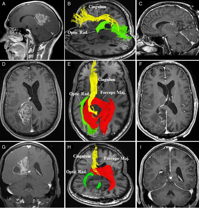
Fig. 5.
(A, sagittal); (D, axial), and (G, coronal) Preoperative MRI with contrast revealed a right-sided enhancing posterior cingulate lesion with an encysted atrium (G). (B, sagittal) and (E, axial) Preoperative high-definition fiber tractography (HDFT) characterized the qualitative changes in the perilesional white matter (cingulum, forceps major [Forceps Maj.] and optic radiations [Optic Rad.]). (H) Postoperative HDFT demonstrated disruption in the forceps major (Forceps Maj.) due to the surgical trajectory through this potentially infiltrated fiber bundle. (C, sagittal); (F, axial) and (I, coronal) Postoperative MRI with contrast demonstrated the resection cavity in the posterior cingulum with resolution of the tumor cyst.

