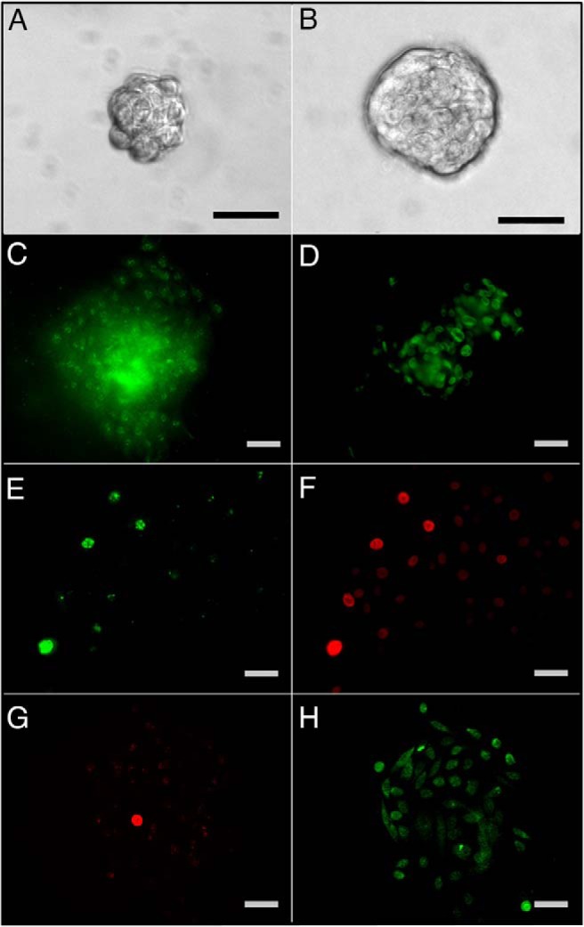
Figure 2.

PS cultured from primary prostate epithelial cells of disease-free organ donors as previously described (11). Day 4 (A) and day 7 (B) PS are clonally generated from a stem-like cell capable of self-renewal and growth through transit amplification of daughter progenitor cells to spheres of 40–50 (day 4) to 100–150 (day 7) progenitor cells at progressing stages of commitment. ERα (C) and ERβ (D) immunostaining of day 7 PS reveals most cells are positive for both receptors. E–H, Primary cells were cultured for 10 days with 1 μM BrdU and transferred to Matrigel culture for sphere formation and BrdU washout. At day 4, PSs were immunostained for BrdU (E and G) to identify the label-retaining stem-like cells. PS were coimmunolabeled for ERβ (F) or ERα (H). E and F, Images for two merged PS reveal that long-term label retaining stem cells (E, green) are strongly ERβ+ (F, red), whereas the daughter progenitor cells have lower relative ERβ protein. G and H, Image of a PS immunostained for BrdU (G, red) and ERα (H, green) shows weak ERα in the stem-like BrdU retaining cell and strong ERα+ in the rapidly dividing progenitor cell population. Scale bar, 50 μM. Panels A–D are reprinted with permission from the authors of another report (11).
