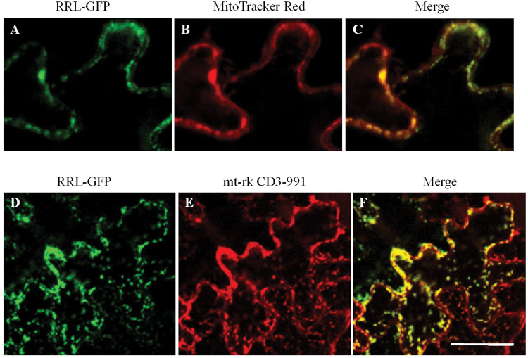
Fig. 1.
Mitochondrial localization of the RRL–GFP fusion protein. (A–C) Subcellular localization of RRL–GFP fusion protein in leaf epidermal cells of Arabidopsis. (A) RRL–GFP, (B) MitoTracker Red staining, (C) co-localization of RRL–GFP and MitoTracker Red. Scale bar=10 μm. (D–F) Subcellular localization of RRL–GFP fusion protein in leaf epidermal cells of tobacco. (D) RRL–GFP, (E) mt-rk CD3-991 (a mitochondrial marker). (F) The overlay image (merge) shows co-localization of RRL–GFP and the mitochondria marker. Scale bar=30 μm. (This figure is available in colour at JXB online.)

