Fig. 3.
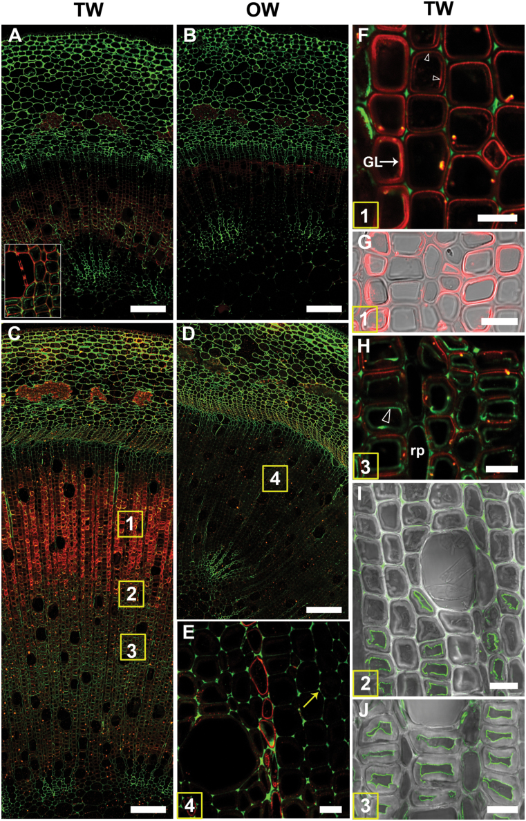
Localization of (1–4)-β-D-galactan (LM5 antibody in red) and homogalacturonan (CCRC-M38 antibody in green) in transverse sections of stems after 1 or 4 weeks of TW induction. Numbers in the squares in (C) and (D) indicate similar areas shown in more detail in subsequent images. (A) to (H) are images of 1 µm resin sections and (I) and (J) are images of 100 µm unembedded sections. (A) Stem after 1 week of tipping showing the distribution of (1–4)-β-D-galactan (red) in TW fibres and of homogalacturonan (green) in the more mature inner side of the stem (detail in the inset). (B) OW side of the same section showing virtually no fluorescence in xylary fibres with the LM5 and CCRC-M38 antibodies. (C) TW side of a 4-week tipped stem showing strong labelling of G-fibres with the LM5 antibody in the outer developing and maturing part of the stem. (D) OW side of the same stem in (C). Fibre cell walls are not labelled or only weakly labelled with the LM5 and CCRC-M38 antibodies. (E) Detail of area 4 in (D). There is no labelling of OW fibre cell walls, indicating the absence of (1–4)-β-D-galactan. Some labelling is seen in ray parenchyma. Homogalacturonan is detected in cell corners (arrow). (F) and (G) Detail of area 1 in (C). LM5 antibody shows differential distribution of (1–4)-β-D-galactan from area 1 in files of G-fibres. The G-layer is labelled in some fibres, whereas in others the interface between the G-layer and SCW is labelled (arrowheads). (H) Detail of area 2 in (C). LM5 is bound mainly to the SCW and CCRC-M38 labels mainly the inner lamella of the G-layer (arrowhead). (I) and (J) Similar areas to 2 and 3 in (C) of unembedded sections showing CCRC-M38 mainly bound to the inner lamella of the G-layer. GL, G-layer; v, xylem vessel; rp, ray parenchyma. Bars: A, B, C, D = 100 µm; E, F, G, H, I, J = 10 µm.

