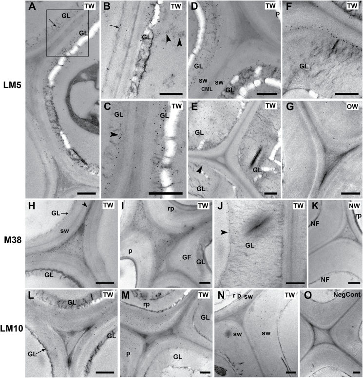
Fig. 7.
TEM immunogold labelling. (A–G) Localization of (1–4)-β-D-galactan with the LM5 antibody in TW (A–F) and OW (G) after 4 weeks of inclined growth. (A) Early G-layer formation. Binding occurs in the G-layer itself or in the interface between the SCW and the G-layer (arrow). The area in the square is magnified in (C). (B) Detail of two adjacent developing G-fibres at early development. In the G-fibre on the left, gold labelling shows that early deposition of (1–4)-β-D-galactan occurs on the inner side of the SCW before the G-layer itself is formed (arrow). The G-layer is already being formed on the adjacent cell and it is labelled. In addition, electron-dense areas in the lumen are also labelled (arrowheads). (C) Detail of the two adjacent fibres in the square in (A) showing differential LM5 labelling occurring in the interface between SCW and the G-layer (arrowhead) or in the G-layer itself. (D) Maturing G-fibres showing thicker G-layers strongly labelled with LM5 antibody next to a ray parenchyma cell with unlabelled SCW. (E) Mature G-fibres showing differential labelling with LM5. Labelling in one of the fibres is mainly in the interface between the SCW and the G-layer (arrowhead). The other two fibres exhibit strong labelling of the G-layer. (F) Fully mature G-fibre. Labelling in these fibres is mainly confined to the SCW-GL interface. (G) OW mature fibres. Very sparse gold labelling is observed in the SCWs of xylary fibres. (H–K) Localization of de-esterified homogalacturonan with the CCRC-M38 antibody in TW after 4 weeks of inclined growth (H–J) and NW in a vertically grown stem (K). (H) Developing G-fibres. Homogalacturonan is localized by the CCRC-M38 antibody in the compound middle lamella (arrowhead) and in the intercellular spaces, which are filled with fibrous material. (I) Homogalacturonan labelling in the intercellular spaces between parenchyma ray cells and G-fibres. (J) Mature G-fibre from the inner part of the stem where CCRC-M38 labelling is mainly confined to the inner lamella of the G-layer (arrowhead). (K) NW fibres walls in a straight stem. CCRC-M38 antibody labelling is present only in cell corners and compound middle lamella. (L–O) Localization of xylan with the LM10 antibody in TW (L–N) after 4 weeks of inclined growth and OW (O). (L) Developing G-fibres. Xylan is restricted to SCWs, but labelling is not very strong. (M) LM10 is bound to the SCWs of ray parenchyma and mature G-fibres. No labelling is detected in the G-layer. (N) OW. Labelling is also confined to the SCWs of fibres and parenchyma cells. (O) Negative control section probed with only the secondary 10nm gold anti-rat antibody. Straight stem fibres showing no gold labelling in any of the cell walls. GF, G-fibre; GL, G-layer; sw, secondary cell wall; rp, ray parenchyma; TW, tension wood; OW, opposite wood; NW, normal wood. Bars: 0.5 µm

