
Figure 2.
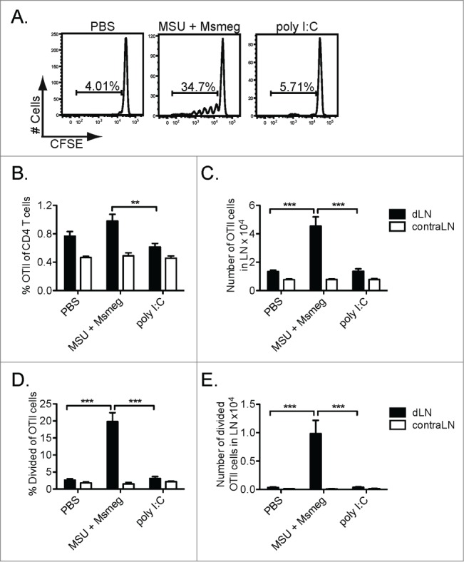
Peri-tumoral treatment with MSU + Msmeg induces the proliferation of tumor-specific CD4+ T cells in dLNs. CFSE-labeled CD45.1+ OTII T cells were adoptively transferred into C57BL/6 mice bearing day 8 B16.OVA tumors. Mice were treated at the tumor site with poly I:C, MSU + Msmeg or PBS vehicle on day 9, and OTII proliferation was assessed in the tumor draining (d) and contralateral (contra) LN on day 14. OTII T cells were identified as single live CD45.1+ CD4+ Vα2+ cells. (A) Representative histograms showing CFSE dilution profiles for OTII cells in tumor-dLN. (B) Frequency and (C) number of OTII cells in LN. (D) Frequency and (E) number of divided OTII cells in LN. Bar graphs show mean + SEM for combined data from 2 independent experiments, each with 5 mice/group.