
Figure 2.
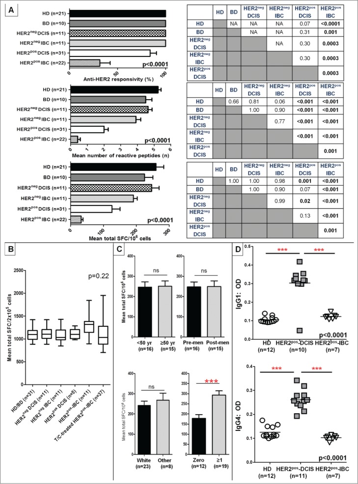
Anti-HER2 CD4+ Th1 responses and IgG1/IgG4 reactivity are progressively lost in HER2pos breast tumorigenesis. (A) IFNγ ELISPOT analysis of PBMCs demonstrated a progressive loss of anti-HER2 CD4+ Th1 response in HER2pos breast tumorigenesis (i.e., HD/BDHER2pos-DCIS HER2pos-IBC), illustrated by anti-HER2 responsivity, response repertoire, and cumulative response (all ANOVA p < 0.001). No differences in Th1 responses were observed between HER2neg-DCIS and HER2neg-IBC (IHC 0/1+) and HD/BD subjects. Post-hoc Scheffé p value comparisons between groups are shown alongside histograms. (B) Anti-CD3/ CD28 stimulus served as positive control for all donors in experiments shown in (A) above; corresponding IFNγ production by ELISPOT in respective patient groups is shown. Results presented as median ± interquartile range (IQR) IFNγ SFC per 2 × 10⁵ cells in box-and-whiskers plots; (C) Variations in anti-HER2 Th1 cumulative responses in HD/BDs donors stratified by donor age (<50 vs. ≥50 years), menopausal status (pre-menopausal vs. post-menopausal) (upper panel); and race (white vs. other), or gravidity (zero vs. ≥1 pregnancies) (bottom panel). Within each Th1 metric, results are expressed as proportion or mean (±SEM). (D) ELISA of serum reactivity against recombinant HER2 extracellular domain revealed significantly elevated anti-HER2 IgG1 and IgG4 antibody levels in HER2pos-DCIS compared with HDs that decayed in HER2pos-IBC patients. ELISA measurements are shown as optical density (OD) at 1:100 sera dilutions (grouped scatter plot, with horizontal line indicating mean OD); (***p < 0.001 by unpaired t-test or ANOVA with post-hoc Scheffé testing, as applicable).