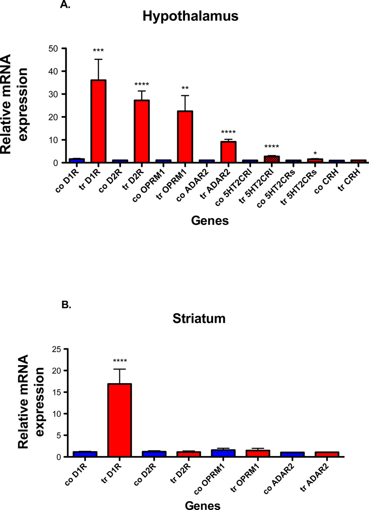
Fig. 2. ADAR2, 5HT2CR, CRH, dopamine and mu receptor mRNA expression in hypothalamus and striatum of control and ADAR2 transgenic mice.
Quantitative real time PCR analyses were done on total RNA using the Bio-Rad SYBR Green assays. (A) The histogram represents the relative mean ±SEM mRNA expression in hypothalamus of ADAR2 transgenic mice (tr) compared to controls (co) (n=8/genotype), (B) the histogram represents the relative mean ±SEM mRNA expression in striatum of ADAR2 transgenic mice (tr) compared to controls (co) (n=4/genotype). ****=p<0.0001, ***=p<0.0005, **=p<0.003 and *=p<0.01.

