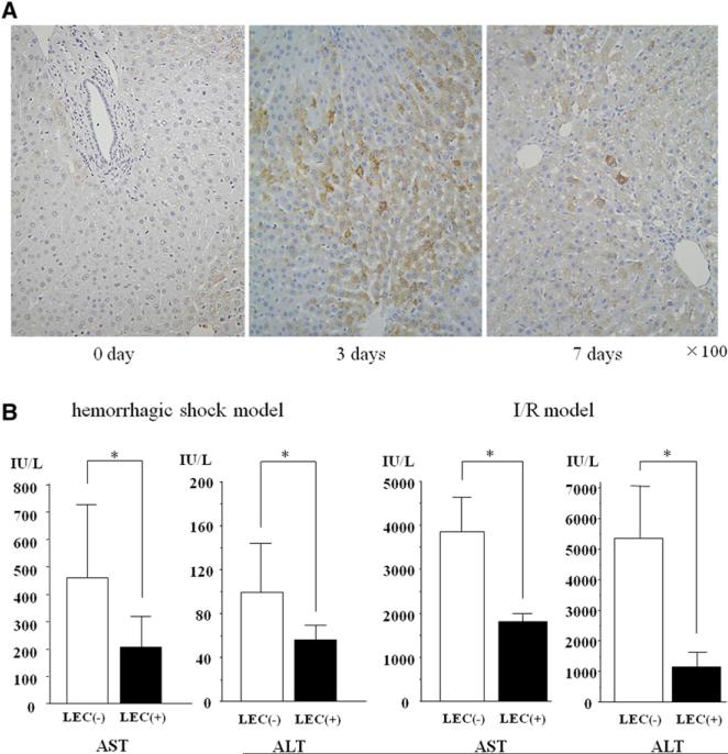
Fig 5.
Induction of endogenous LECs in the entire liver using SST/PTO model prevented liver injury after hemorrhagic shock and I/R. A, Liver tissues were collected and fixed at 3 or 7 days after portal trunk ligation. Liver sections were stained for AFP to identify LECs. Representative sections showed that AFP staining was increased after PTO. The population of LECs was 58.3 ± 7.31 and 9.8 ± 6.37 cells per 1,000 hepatocytes on days 3 and 7, respectively. Original magnification, ×100. B, Increases of serum AST and ALT levels were suppressed in LEC-induced liver 24 hours after resuscitation in hemorrhage shock model and 3 hours after reperfusion in I/R model (*P < .05 versus control, n = 6 per group). LEC([−]): SST-subjected group, LEC(+): STT/PTO-subjected group. (Color version of figure is available online.)

