. 2015 Sep 30;10(9):e0138917. doi: 10.1371/journal.pone.0138917

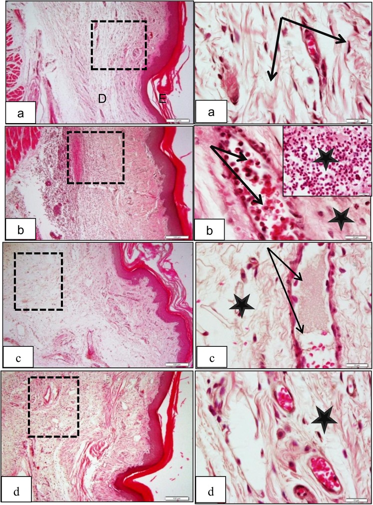
Fig 7. Low and magnified power of rat paw sections stained by H&E, (a) Control group, E: epidermis, D: dermis with no signs of vascular congestion or inflammatory cells (dotted square) with normal capillaries (black arrows) and connective tissue dermis (stars). (b) Carrageenan group, showing marked inflammatory infiltration in the deep dermis (D), capillary dilation and congestion with neutrophils margination prior to escape into the surrounding tissue (arrows;) (c) Xestospongia testudinaria methanolic extract group, showing significant decrease of the inflammatory cells in the dermis and within blood vessels. (d) Indomethacin group, showing a decrease in both inflammatory cells infiltration and vascular congestion.

Fig 7